MORE POSTS
Surgical Management of the Charcot Ankle2024-01-17
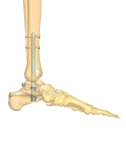
Charcot neuroarthropathy of the ankle is an especially challenging condition that often leads to instability and destruction of the joint with significant morbidity requiring an amputation in many instances1-3. According to Brodsky et al., the ankle is affected in 9% of patients with Charcot neuroarthropathy, whereas the hindfoot is involved in 30-35%3. The overall goal of Charcot ankle reconstruction is to achieve a painless, plantigrade and stable foot, and thus avoid ulceration with subsequent infection1,2. However, this procedure is also associated with a high complication rate1,2.
Postoperative Management in Charcot Foot Reconstruction2024-01-10
The successful treatment of Charcot foot does not end with a completed surgery; success also requires a diligent postoperative care regime. This devastating condition mainly affects patients with diabetes and peripheral neuropathy, resulting in bone destruction and joint deformation1. During the initial, inflammatory stage, the recommended treatment consists of off-loading with total contact casts or custom orthotics to prevent further damage, although surgical management is indicated in cases of severe instability or soft tissue compromise2. After the acute stage, the focus of treatment is on deformity correction and foot reconstruction to prevent further damage and improve the patients’ quality of life2.
Foot and Ankle Charcot Reconstruction with External Fixation2023-12-20
Surgical reconstruction of foot and ankle Charcot with external fixation is especially useful in the presence of poor bone quality, ulceration, compromised soft tissue, active infection, or osteomyelitis – cases in which the use of internal fixation is limited or contraindicated1.
Charcot Foot Reconstruction Using Internal Fixation2023-12-12
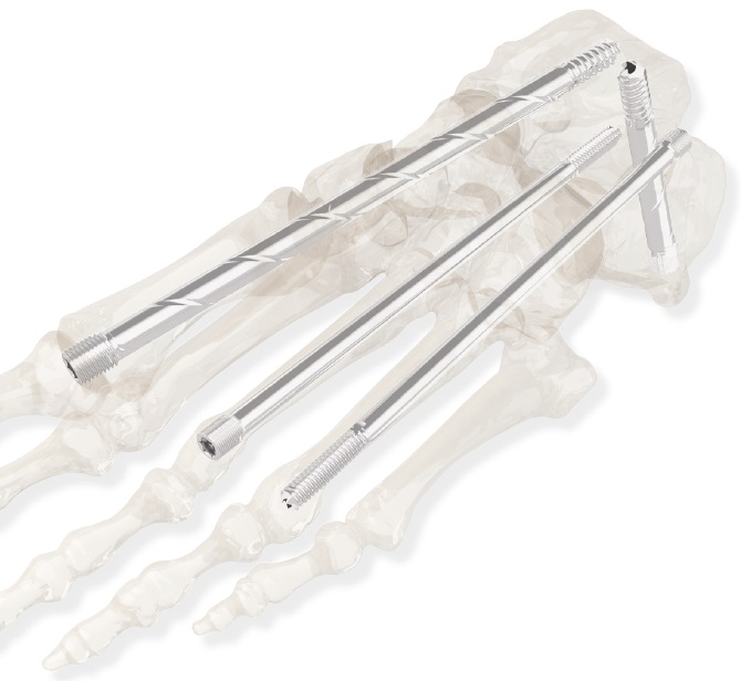
Appropriate timing for surgical intervention is a matter of controversy among specialists1. While in the past Charcot foot surgical reconstruction was mainly reserved to salvage the limb, today surgeons also perform operative corrections to ensure patients can use commercially available footwear instead of cumbersome orthoses1. Regardless, the main goal of Charcot foot surgery, independent of the fixation method, is to create a stable and plantigrade foot. This article will discuss different approaches for Charcot foot surgery with internal fixation, focusing on midfoot reconstruction.
Charcot Arthropathy Symptoms and Initial Stage2023-11-21
Charcot arthropathy occurs in the presence of peripheral neuropathy and metabolic abnormalities. Today it is seen more commonly in patients with long-standing diabetes1,2. Its pathogenesis, although still largely unknown, involves several pro-inflammatory pathways, which stimulate osteoclastic activity3.
Medical Resources for Charcot Foot & Ankle Specialists2023-11-14
There is an overwhelming wealth of scientific literature available on Charcot foot. In this article, we gather and summarize some of the most interesting sources, including Charcot neuroarthropathy of the foot and ankle reviews, diabetic foot reviews, and diabetic foot care articles.
The Experts’ Definition of Diabetic Charcot Foot2023-11-14
Charcot foot is a progressive, devastating pathology defined by bone and joint changes that occur as a result of loss of sensation. Also called Charcot joint or neuropathic arthropathy, this condition is almost always associated with the vascular and neurological changes caused by poorly controlled diabetes1.
Charcot Surgical Reconstruction2023-11-14
Charcot foot, also called Charcot neuropathy, is a condition caused by a combination of peripheral neuropathy and vascular disorders that results in progressive joint damage and eventually foot instability1. If left untreated, the consequences for patients are devastating, with reduced quality of life, inability to weight-bear, and an increased risk of amputation2.
Surgical Approaches for Charcot Foot2023-11-14
Charcot neuroarthropathy is a rare, but devastating complication of diabetes1. Progressive destruction of the bones and joints of the foot cause deformities and thus alter foot biomechanics1. If not recognized nor treated properly, the disease can result in ulcer formation, infection, and progression of the disease with joint instability and bone destruction2,3. In the worst case, limb amputation is required4,5.
Post-Operative Management of Deformity Correction Surgery with External Fixation2023-11-09
The successful outcome of a deformity correction with external fixation surgery relies also on a meticulous post-operative management approach. It is critical to optimal healing and the restoration of functionality. Wound care, pain control, rehabilitation exercises, monitoring of complications, and regular follow-ups are integral to a faster recovery.[1]
Why Playing Video Games Can Be Good For Patient’s Health?2023-11-08
Modern healthcare is evolving fast and creatively. One of the key difficulties faced by a young child who has undergone orthopedic surgery is to contain their movement to allow for appropriate healing. The younger the child, the more challenging it is to remain immobile.
The Growing Skeleton and Deformity2023-11-02
The human skeleton consists of an intricate architecture of bones – a living tissue that is perpetually undergoing transformation throughout our lives. The most significant portion of bone development, repair, and remodeling occurs during a person's growth period in their youth and adolescence, contributing to our adult body structure. However, certain factors can disrupt this process, leading to skeletal issues like limb deformity.[1]
Mobile Apps in Orthopedic Post-surgery2023-10-19
Modern healthcare is taking orthopedic aftercare to the next level by ensuring a more personal touch to the patient, once they are released from their hospital stay. Going home after any minor or major surgery is a time of heightened anxiety and proper post-operative (post-op) care instructions and resources are key to ensure that the patient can benefit most from the treatment.
Ultrasound and Cementless Prosthesis Revision2023-09-15
Total joint replacement surgeries are one of the most common procedures practiced in orthopedics with hip and knee replacements being the most common procedures performed, followed by shoulder and elbow replacements. Over time, a total joint replacement begins to wear and tear, and a revision procedure may be necessary to replace worn-out joint components with a new or improved prosthesis.
Ultrasound and the Removal of Bone Cement in Revision Arthroplasty Surgery2023-09-12
Revision arthroplasty is a procedure performed when a worn-out hip, knee, shoulder, or elbow joint replacement component requires replacement with a new prosthesis. Hip and knee arthroplasties are some of the most common procedures undertaken today. It is estimated that in 10 years, the number of primary total hip arthroplasty interventions will grow by 71% to 635,000 procedures, and primary total knee arthroplasties will undergo an 85% increase, totaling 1.26 million procedures in the United States. Consequently, total hip and total knee revisions are projected to grow by 137% and 601%, respectively, between 2005 and 2030.[1] Shoulder arthroplasty is also a prevalent joint replacement surgery, being the third most common after hip and knee replacement. In terms of complications, total elbow arthroplasty has complication rates higher than that of total joint arthroplasty of all other joints.
The Challenges Associated with Revision Arthroplasty2023-09-07
Painful arthritic and inflammatory conditions of the joints are common and often lead to the need for surgical intervention. Severe cases require complete joint replacement such as total knee arthroplasty, total hip replacement and total shoulder or elbow arthroplasty.
Principles of Multiplanar Deformity Analysis and Correction in Long Bone2022-02-17
A multiplanar deformity analysis can be both simple and complex: a correct determination of angular and metric deformity apex and plane is a fundamental task. This is true in both the preoperative surgery planning and the postoperative management phases, in order to obtain the most accurate correction and promote the quickest recovery for the patient’s well-being.
Calculation of the True Plane of the Deformity2022-02-17
The initial assessment of any lower extremity deformity is fundamental to plan a successful surgical program of correction, as well as an effective post-operative management of the healing process. The assessment should include the X-rays of full length weight bearing of both legs, with a complete axis evaluation to fix the position of the deformities, which is “the true plane of the deformity”.
Tips for Timely Diagnosis of Charcot Foot and Ankle2022-01-17
It is known that around 9 per cent of patients with diabetic neuropathy and foot ulcers develop a Charcot foot and ankle. This pathology often goes unrecognized, particularly during the acute phase, until severe complications occur, such as an abnormal pressure distribution on the plantar fascia with bones weakened enough to fracture, followed by a marked foot and ankle deformation and, in the worst cases, by amputation. Thus, today, one of the most crucial questions is how to timely diagnose this condition.
Acute or Gradual Correction of a Deformity?2022-01-17
Although several studies have reported good results of the treatment of the lower extremity deformity combined with shortening – a condition that often means disabling impairment in the daily life activities – to date there are still very few studies that review the evidence by comparing the outcomes of acute versus gradual correction.
Magnetic Resonance for Charcot Foot and Ankle Diagnosis2021-12-21
Charcot Foot and Ankle is a significant problem caused by peripheral neuropathy, with an estimated incidence of 0.3 to 7.5% in the diabetic population. This condition, which is also known as Charcot arthropathy, Charcot joint, or neuropathic arthropathy, affects the soft tissues, joints, and bones in the foot and ankle. If left untreated, it may lead to progressive foot deformity, ulceration, or osteomyelitis. In the most advanced stages, it may be necessary to resort to limb amputation.
Tips for the Prevention of Charcot Foot and Ankle2021-12-17
Charcot Foot and Ankle is a severe condition that, if left untreated, may lead to deformity, permanent disability, amputation or ultimately to the patient’s death. People with diabetic neuropathy are at high risk of developing Charcot Foot and Ankle, because of the decreased sensation and diminished ability to feel temperature, pain or trauma.
CORA (Center Of Rotation of Angulation)2021-12-17
The term CORA (Center of Rotation of Angulation) means a method of mechanical and anatomical axis planning based on joint orientation, which has gained international recognition and acceptance. Many surgeons from all over the world apply this method to limb lengthening and deformity correction, and the CORA principles and concepts have clearly shaped both design and functionality of the last generation of external devices.
Computerized Tomography for Charcot Foot and Ankle Diagnosis2021-12-14
Charcot Foot and Ankle, also known as Charcot arthropathy or Charcot joint, is a condition caused by peripheral neuropathy, leading to the progressive degeneration and destruction of the soft tissue, joint, and bone. In modern society, the most common cause for this neuropathic neuropathy is diabetes mellitus, with long-term diabetic patients having an increased risk of developing the condition.
Radiography for Charcot Foot and Ankle Diagnosis2021-12-07
Charcot Foot and Ankle, also known as Charcot arthropathy, neuropathic arthropathy or Charcot joint, is a progressive condition that affects the bones, joints and soft tissue of the foot and ankle. It is caused by peripheral neuropathy. Today, diabetes is the most common cause for developing Charcot Foot and Ankle.
Charcot Foot and Ankle Causes: Where the Pathology Starts2021-11-25
Charcot Foot and Ankle, also known as Charcot arthropathy, is a progressive, inflammatory condition that affects the bones, joints, and soft tissues in the feet of people with peripheral neuropathy. If left untreated, Charcot of the foot and ankle leads to severe deformity, ulceration, and – worst-case scenario – to limb amputation.
Weight Bearing and Dynamization2021-11-17
Weigh bearing is considered a significant clinical issue particularly when an external fixator has been applied to a lower limb. If mechanical stability is in fact a mandatory condition for promoting fracture healing, other key components for a full recovery of the bone are early weight bearing and dynamization.
The Compression-Distraction Technique2021-11-17
In the compression-distraction technique, one site of the bone is lengthened and another site is immediately or gradually compressed. In the lower limb, the compression-distraction technique is generally performed in the presence of leg shortening associated with small bone defects – less than 3cm in the tibia and/or less than 5cm in the femur.
Charcot Foot and Ankle: Definition and Classification2021-11-12
Charcot Neuroarthropathy, also called Charcot Foot and Ankle, is a progressive and inflammatory condition that weakens the soft tissues, bones, and joints of the foot and ankle. This complex and challenging type of neuropathic arthropathy is commonly linked to diabetes. If left untreated, it can lead to devastating consequences, such as deformity, ulceration, and infection. In the worst-case scenario, it may result in limb amputation.
Dealing With Pin Site Problems2021-10-17
To prevent and manage pin site problems, the patient should be clearly informed of the protocol he/she needs to follow for pin site care in the post-operative phase. And not only the patient, but all people involved in the healing process outside the hospital should know the protocol.
Controversies on the Role of Dynamization in the Bone Healing Process2021-10-17
Dynamization (dynamic loading and controlled motion at the fracture site) is considered a controversial and evolving issue, besides the positive functional results observed both in experimental studies based on animal models, and in human clinical cases in the last 30 years. Currently one crucial matter is to understand the biomechanical, biological and physiological effects of dynamization on the bone healing process, in order to show its safety and effectiveness.
The Reverse Planning Method (RPM)2021-05-27
When an orthopedic surgeon has to perform a bone deformity correction, he or she may choose to apply an extrinsic method of correction such us an external fixator, or an intrinsic one, such us an intramedullary nail. In both cases, a surgeon will always have to consider the patient’s bone quality through three dimensional parameters: length, alignment and torsion, and accurately the entire 3D correction process. If using an intramedullary nail, an effective planning method is the Reverse Planning Method (RPM) developed by Prof. Rainer Baumgart in 2009.(1)
The Surgical Approach to the Charcot Foot and Ankle Treatment2021-05-24
Charcot Foot and Ankle is a complex, challenging and sometimes devastating inflammatory foot disease which causes loss of sensation, weakening of the bones and collapse of the weight bearing joints. Most often due to diabetes and underlying neuropathic arthropathy, it needs a multidisciplinary team approach in order to treat it properly and prevent amputation.
The Orthofix Philosophy of Pediatric Solutions2021-05-23
A child may need the help of a pediatric orthopedic surgeon for many reasons: musculoskeletal injuries caused by playing a sport, jumping, climbing or cycling, and conditions that have to be treated by a specialized team. In addition to several pediatric solutions and a deep knowledge within the orthopedic surgical field, Orthofix offers a complete and effective treatment philosophy.
Internal Fixation vs External Fixation2021-05-22
A typical question that a surgeon has to face when deciding the treatment of a bony injury is when to use internal fixation instead of external fixation. A common answer is “when the patient is compliant, with sufficient bone and soft tissue quality, and presents less complex fractures”. Surgeon’s expertise and preference may also affect the final surgical plan.
Basic Considerations of Deformity Correction: Anatomical and Mechanical Axis2021-05-15
Bone deformity is a three-dimensional issue, considering the three-dimensional shape of bones and joints. All basic considerations of deformity correction by using an external device should include the fundamental concepts of Anatomical and Mechanical Axis. These concepts allow a better understanding of the effects of adjustments or modifications in one plane when managing a three-dimensional problem such as bone deformity.
The Multifocal Procedure with the Limb Reconstruction System (LRS)2021-05-14
The multifocal procedure, in which two osteotomies are performed, is indicated in the presence of large bone defects, extreme shortening, shortening combined with metaphyseal deformity where lengthening takes place in more than one site, and fractures with soft tissue defects and exposed bone. The application technique of the external device is quite similar to that for bone transport, with the only difference being that in this procedure two compression-distraction units are used simultaneously.
The Monofocal Procedure with the Limb Reconstruction System (LRS)2021-05-13
The monofocal procedure is performed for lengthening at a single osteotomy site, for deformity correction with or without lengthening, and when increased stability is required. This procedure may be also used in bone and soft tissue defects when the bone loss is less than 2cm.
The Bone Transport Technique2021-05-11
The bone transport technique allows regeneration of the bony tissue in the patient’s limb, in those cases associated with significant bone loss or defect – such as acute fractures, aseptic or infected nonunions, chronic bone infections, tumors, osteomyelitis. Bony regeneration is generally used to fill a gap or missing bone.
Key Points for Healthy Pin Sites2021-05-08
Preserving healthy pin sites is essential to effective external fixation, for the prevention of pin track infection and related complications. All people involved in the healing process – general practitioner, surgeon, nursing staff, patient and family – play a fundamental role in order to achieve the best result. Here are some useful key points.
What Is Arthrodiatasis (Articulated Joint Distraction)?2021-05-07
The term arthrodiatasis was first coined in 1979 in Verona, Italy. It comes from Greek, meaning “joint-through-to stretch out”, in a few words articulated distraction of the joint. It is a technique exclusively referring to external fixation, which was developed initially for the conservative treatment of the osteoarthritis of the hip, and later successfully used to cure other conditions.
What Is Arthrodesis (Joint Fusion)?2021-05-06
Arthrodesis may be defined as the surgical immobilization of a joint by fusion of the bones. It is commonly referred to as joint fusion, also known as artificial ankylosis or syndesis. It can be performed in several parts of the body, and it is indicated in the presence of painful joints, often due to arthritis or osteoarthritis, with highly compromised mobility.
The Orthofix Philosophy2021-05-05
“Respect for, and enhancement of the natural physiological process of fracture healing” may be considered the philosophy which has inspired the research and design of all products developed by Orthofix in the last 40 years, starting with the set of devices belonging to the internal and external fixation system.
Orthofix Dynamization and Initial Devices2021-05-03
The Orthofix history began at the end of the 70s in Italy with the work of orthopedic researcher Giovanni De Bastiani. He proposed the concept of “dynamization” and developed a modular external fixation device designed to allow the release of axial forces at the proper time during the healing process, thus transferring a progressive load to the fracture site. After this first device, other devices were developed by Orthofix to better satisfy patients’ and doctors’ needs.
The Concept of Dynamization2021-05-02
Stability does not mean rigidity. Medical philosophy has evolved from the aim to eliminate micromotion between the bony fragments to the idea of controlling it. Micromotion is necessary for external callus formation, and therefore for a functional bone healing. External fixation offers the opportunity to control both micromovement and load at the fracture site, which corresponds to the concept of dynamization.
Bone Healing and The Role of External Fixation2021-05-01
When a bone breaks, all the systems work for the bone healing. Fracture healing is a natural, physiological process, and the surgeon may help nature to restore the biological, structural and mechanical properties of the bone as they were before the break, transforming the fracture into a healed, normal and straight bone.