Skip to the content
Abs
  • Intro
  • Company
  • Blog
  • Mobile app

THIS INFORMATION IS FOR HEALTHCARE PROFESSIONALS ONLY

  • Intro
  • Company
  • Blog
  • Mobile app

Search

Blog

Would you like to access to an extensive range of learning and discussion opportunities? SUBSCRIBE TO THE ORTHOFIX ACADEMY™

Categories

  • Blog

MOST READ

  • 2022-02-17Principles of multiplanar deformity analysis and correction in long bone

  • 2022-02-17Calculation of the True Plane of the Deformity

  • 2022-01-17Tips for timely diagnosis of Charcot foot and ankle

  • 2022-01-17Acute or gradual correction of a deformity?

  • Tags charcot_foot_and_ankle
Computerized Tomography for Charcot Foot and Ankle Diagnosis
2021-12-14

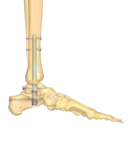
Charcot Foot and Ankle, also known as Charcot arthropathy or Charcot joint, is a condition caused by peripheral neuropathy, leading to the progressive degeneration and destruction of the soft tissue, joint, and bone. In modern society, the most common cause for this neuropathic neuropathy is diabetes mellitus, with long-term diabetic patients having an increased risk of developing the condition.

Given its progressive nature, a late Charcot Foot Diagnosis can lead to considerable morbidity, including potential limb loss. However, the clinical and radiological diagnosis of Charcot Foot and Ankle are both challenging. Furthermore, the clinical presentation of acute Charcot neuropathy and diabetic osteomyelitis is similar, and both conditions can co-exist in the same extremity, adding to the complexity of Charcot Foot Diagnosis.

Although clinical history, risk factors, and physical and lab exams are important in the diagnosis of Charcot Foot and Ankle, imaging tools play a decisive role in arriving at the definitive diagnosis and adequate treatment. In this article, we will focus on the role of Computerized Tomography (CT) imaging in the diagnosis and management of Charcot arthropathy.

How to use CT scans to diagnose Charcot Foot and Ankle

Although the use of CT scans in neuropathic arthropathy is not well investigated, this imaging tool has higher sensitivity for early intra-articular fractures that are not readily visualized in plain radiographs. CT can also show periosteal new bone formation and small foci of gas within bone in a better way than MRI can. Other findings that can be evidenced with the use of CT are cortical destruction, anatomical deformities, structural bone damage, fractures, subluxations, and bone callus formation.

When MRIs are contraindicated, the use of CT is particularly useful to visualize all bones of the foot, particularly the midfoot, which is difficult to see well on radiography and one of the crucial points of damage in Charcot Foot and Ankle.

Currently, the most used CT scanning technique is the multidetector computed tomography (MDCT), and its use with reconstruction and three-dimensional (3D) computer-generated images from multiple planes is advantageous in preoperative planning and follow-up of immobilization therapy.

When using the MDCT to create 3D images, both feet should be scanned to determine bilateral deformities and differences between feet. To determine if there is subluxation of the tarsometatarsal joints, radiologists should evaluate the transverse and coronal planes of the foot.

In early stages, when Charcot Foot Symptoms are easily mistaken with other conditions and radiological signs can be non-existent or very subtle, the use of MDCT enables the analysis of normal bone density, joint distension, existence of bony debris, joint disorganization and dislocation, collectively known as the descriptive “five D’s” of neuropathic arthropathy.

The patients’ role

Early diagnosis of Charcot Foot and Ankle may be difficult, as the presentation with swelling and erythema can be overlooked or mistaken with other conditions, such as cellulitis or erysipelas. The use of 3D CT scans can help radiologists to address the spatial orientation of the bones and joints, especially the subtle but complex midfoot and rearfoot joint alterations in early stages of Charcot arthropathy.

Since the alterations caused by Charcot Foot and Ankle may be initially elusive, it is important that they are detected as soon as possible. To accomplish this, the patient must be aware of his or her crucial role in the disease management by doing regular check-ups, carefully taking care of their feet, and promptly communicating any alteration to a member of their multidisciplinary healthcare team.

Failing to take preventative measures and delaying treatment can result in burdensome complications, such as pronounced foot deformity, ulcers, infection, and ultimately lead to limb amputation.

If you wish to help your patients know more about Charcot Foot and Ankle, its link with diabetes, and their key role in the management of their disease, we suggest downloading the Patient Informative Cards “Stand On Your Feet: A Program of Empowerment for Your Diabetic Foot Management.”

References

  • Mautone M, Naidoo P. J Med Imaging Radiat Oncol 2015 Aug;59(4):395-402.
  • Marmolejo VS et al. Am Fam Physician. 2018 May 1;97(9):594-599.
  • Schoots et al Semin Musculoskelet Radiol. 2010 Sep;14(3):365-76.
  • Rogers LC, Bevilacqua NJ. Clin Podiatr Med Surg 2008; 25: 263–274, vii.
  • Ergen FB et al. Diabet Foot Ankle. 2013 Nov 20;4.
< Calculation of the True Plane of the Deformity

MORE POSTS

Tips for timely diagnosis of Charcot foot and ankle2022-01-17

It is known that around 9 per cent of patients with diabetic neuropathy and foot ulcers develop a Charcot foot and ankle. This pathology often goes unrecognized, particularly during the acute phase, until severe complications occur, such as an abnormal pressure distribution on the plantar fascia with bones weakened enough to fracture, followed by a marked foot and ankle deformation and, in the worst cases, by amputation. Thus, today, one of the most crucial questions is how to timely diagnose this condition.

Magnetic Resonance for Charcot Foot and Ankle Diagnosis2021-12-21

Charcot Foot and Ankle is a significant problem caused by peripheral neuropathy, with an estimated incidence of 0.3 to 7.5% in the diabetic population. This condition, which is also known as Charcot arthropathy, Charcot joint, or neuropathic arthropathy, affects the soft tissues, joints, and bones in the foot and ankle. If left untreated, it may lead to progressive foot deformity, ulceration, or osteomyelitis. In the most advanced stages, it may be necessary to resort to limb amputation.

Tips for the prevention of Charcot foot and ankle2021-12-17

Charcot foot and ankle is a severe condition that, if left untreated, may lead to deformity, permanent disability, amputation or ultimately to the patient’s death. People with diabetic neuropathy are at high risk of developing Charcot foot and ankle, because of the decreased sensation and diminished ability to feel temperature, pain or trauma.

Radionuclide for Charcot Foot and Ankle Diagnosis2021-12-09

Early Charcot Foot Diagnosis is essential to a proper management, but this can be challenging. There are some factors that should increase clinical suspicion for acute Charcot arthropathy, namely if the patient presents with:

Radiography for Charcot Foot and Ankle Diagnosis2021-12-07

Charcot Foot and Ankle, also known as Charcot arthropathy, neuropathic arthropathy or Charcot joint, is a progressive condition that affects the bones, joints and soft tissue of the foot and ankle. It is caused by peripheral neuropathy. Today, diabetes is the most common cause for developing Charcot Foot and Ankle.

Charcot Foot and Ankle Diagnosis: The Multidisciplinary Approach2021-12-02

Patients with Charcot neuroarthropathy, a condition that affects the bones, joints, and soft tissues of the foot and ankle, greatly benefit from a timely diagnosis and appropriate treatment. However, Charcot Foot and Ankle diagnosis can be quite challenging, especially in an early stage, where acute symptoms can be similar to other conditions.

Charcot Foot and Ankle: Patient Questions and Doctor Answers2021-11-30

Linked to peripheral neuropathy, Charcot Foot and Ankle is one of the serious foot complications affecting people with diabetes. Foot injuries that go unnoticed due to the reduced pain sensation accumulate, and, over time, there is bone deformation, ulceration, and infection. In a worst-case scenario, amputation may be necessary.

Charcot Foot and Ankle causes: Where the Pathology Starts2021-11-25

Charcot Foot and Ankle, also known as Charcot arthropathy, is a progressive, inflammatory condition that affects the bones, joints, and soft tissues in the feet of people with peripheral neuropathy. If left untreated, Charcot of the foot and ankle leads to severe deformity, ulceration, and – worst-case scenario – to limb amputation.

The Stages of Charcot Foot and Ankle Disease and the Importance of a Quick Diagnosis2021-11-23

When dealing with Charcot Foot Disease, a swift diagnosis is crucial to improve patient outcomes and avoid complications such as foot deformity, ulceration, and in a worst-case scenario, limb amputation. However, not all physicians are able to recognize the initial signs of a Charcot Foot and Ankle in their diabetic patients.

Emergency in Diabetic Foot and Ankle2021-11-16

Diabetic foot is one of the most important sources of morbidity in patients with diabetes, affecting 6.4% of patients worldwide. It is the most common cause of lower extremity amputation.

Charcot Foot and Ankle: Definition and Classification2021-11-12

Charcot Neuroarthropathy, also called Charcot Foot and Ankle, is a progressive and inflammatory condition that weakens the soft tissues, bones, and joints of the foot and ankle. This complex and challenging type of neuropathic arthropathy is commonly linked to diabetes. If left untreated, it can lead to devastating consequences, such as deformity, ulceration, and infection. In the worst-case scenario, it may result in limb amputation.

The surgical approach to the Charcot foot and ankle treatment2021-05-24

Charcot foot and ankle is a complex, challenging and sometimes devastating inflammatory foot disease which causes loss of sensation, weakening of the bones and collapse of the weight bearing joints. Most often due to diabetes and underlying neuropathic arthropathy, it needs a multidisciplinary team approach in order to treat it properly and prevent amputation.

Orthofix

2021 © Orthofix Srl
Via delle Nazioni 9
37012 Bussolengo, Verona - Italy
Phone: +39 045 6719000
Fax: +39 045 6719080
Email: customerservice@orthofix.it

THIS INFORMATION IS FOR HEALTHCARE PROFESSIONALS ONLY

This website is for educational purposes only and is not a substitute for a consultation with a physician or healthcare professional. The information provided herein is a service, should be viewed only as opinion. The information contained herein has been obtained from sources deemed ethical and reliable, but completeness is not guaranteed.

LinkedIn YouTube

CALL US
+39 045 67 19 000

EMAIL US
customerservice@orthofix.it

Categories

ORTHOFIX CORPORATE WEBSITE PRIVACY POLICY

COOKIE POLICY

TERMS OF USE

2013 © Orthofix Srl

Copyright © Orthofix. All rights reserved.

Top ↑ Top ↑

Some materials may not be available in your Country. Please contact your sales representatives for availability.

RESERVED ACCESS

This website is reserved only for Healthcare Professionals and their staff. Therefore, the access to these information is denied to non professional audience.

This website uses third-party cookies to improve your experience. Find out more and set your cookie preferences here.
By continuing to use our site or clicking "I accept" you consent to use our cookies.
I accept