


To prevent and manage pin site problems, the patient should be clearly informed of the protocol he/she needs to follow for pin site care in the post-operative phase. And not only the patient, but all people involved in the healing process outside the hospital should know the protocol.
Pin sites should heal around the wires and pins, like a pierced ear heals.(1)
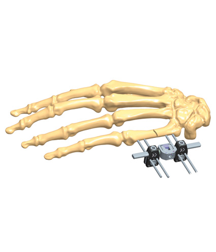
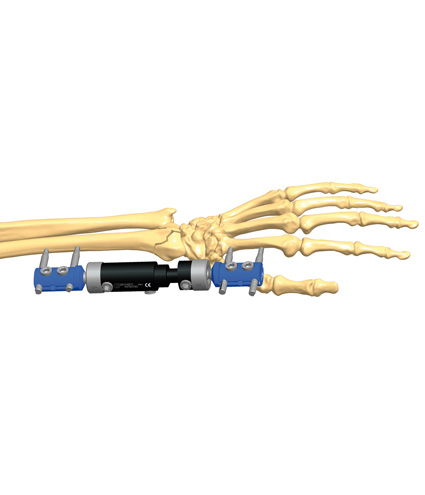
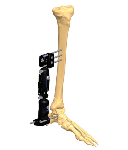
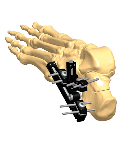
Methods and techniques proposed to deal with pin site problems, associated with the use of external fixation, are varied and constantly evolving. The standard Orthofix guidelines are based on the Checketts-Otterburn method for pin site care, and their system of classification of pin site infections, which divides pin site infections into two groups: minor (grade 1-3) and major (grade 4-6).(2)
Pin site irritation and/or infection can be resolved without consequences if dealt with on time by using proper pin site care, which implies a correct pin site hygiene, associated with antibiotic therapy when necessary. If not properly managed, pin track infection may decrease the stability of the pin-bone interface, as well as provoke screw loosening.
Loss of serous fluid is a common problem, more frequent in obese patients and with femoral screws, and it should not be confused with an infection. This is generally due to the excessive patient mobility: in this case, a correct application of the pin site care protocol is all that is needed.
An infection starts with an inflammatory state of the pin track – the exudate is purulent and the skin around the screw is red, warm and swollen. Although pin track infection is quite common, it seldom leads to major complications, with more than 90% of infections responding well to local or systemic antibiotic management.(3)
Pin track care should be started as soon as the pin site infection is identified, after taking a bacterial swab and before the targeted antibiotic treatment. This includes cleaning of the pin-skin interface with a chlorhexidine solution and absorbent dressing in the presence of excessive exudate.
If the infection is in the lower limb, weight bearing should be restricted until the infection is solved. If the patient’s conditions do not improve, he or she should return to hospital for more intense therapy, and the possible removal of the screws or wires involved.
During the pre-dynamization phase, if X-rays show signs of osteolysis around the screw, with evidence of screw loosening, it is recommended to reposition the screw, when simple removal would compromise the frame’s stability. In the case of osteolysis occurring during the early dynamization phase it may be necessary to remove both fixator and screws, and apply a functional brace. When osteolysis occurs during the late dynamization phase, it is possible to leave the device in place until final healing is reached, constantly monitoring the entire situation.
References
- Rogers LC, Bevilacqua NJ et al. 2007. Predictors of postoperative complications of Ilizarov external ring fixator in the foot and ankle. J Foot Ankle Surg; 46(5):372-5.
- Checketts RG, MacEachern AG, Otternburn M 2000. Pin track infection and the principles of pin site care, in De Bastiani G, Apley AG and Goldberg AAJ (eds). Orthofix External Fixation in Trauma and Orthopaedics. London: Springer; 97-103.
- Schalomon J, Petnehazy T et al 2007. Pin track infection with external fixation of pediatric fractures. J Ped Surg; 42:1584-7.


