
Large bone defects are mainly caused by trauma, infection, or tumors, and may be accompanied by soft tissue damage.(1,2) Bone reconstruction in these cases must be done by means of orthopedic surgery, since large bone gaps will not heal spontaneously.(1,3) There are several options to reconstruct bone defects, including bone grafting, the induced membrane technique, and bone transport.(1,3)
The bone transport technique was developed by Professor Gavriil Ilizarov in 1951, providing a solution to address a region where bone is or will be missing after resection.(4) The key principle of this technique is the “law of tension stress”, which describes how continuous, slow traction can promote new bone growth due to its natural regeneration and plasticity.(4,5) This advanced surgical approach, although possessing a steep learning curve, has the advantage of addressing several problems besides the bone defect, including malalignment and soft tissue loss.(2,6) There is extensive scientific evidence regarding the use of the bone transport technique in patients with severe and complex bone injuries, which highlights its importance in limb salvage procedures and to avoid amputation.(7,8)
What is the Bone Technique?
This surgical technique allows for new bone growth in an area where there is a bone gap. When the bone is broken in a controlled way, new bone is formed in the gap between the existing bone ends. While it is being formed, the new bone is pliable and keeps developing while the bone ends are slowly pulled apart.(4) This is the foundation of distraction osteogenesis, a process also used in bone lengthening procedures.(9,10) In the bone transport technique, a healthy bone segment is moved into the area of the bone gap along with its soft tissue envelope, allowing for new bone to grow in the area that is being distracted.(1,3,11) Meanwhile, the proximal and distal bone segments maintain their alignment and stability.(1) When the advancing bone makes contact with the other bone end, the bone ends are then compressed together to promote union and callus formation at the docking site.(9,10)
The main goals of the bone transport technique are to restore limb function and strength while maintaining its structural integrity in cases of bone loss.(8) Although other techniques, such as bone grafting, can also be effective in certain situations, they usually do not address instability or bone deformity at the docking site.(12)
Historically, amputations were often employed in cases of trauma with significant bone loss.(2) Although they are still employed in extreme cases of mangled extremities, modern limb salvage procedures, pioneered by Prof. Ilizarov and his bone transport technique, have decreased the need for amputations.(2,7)
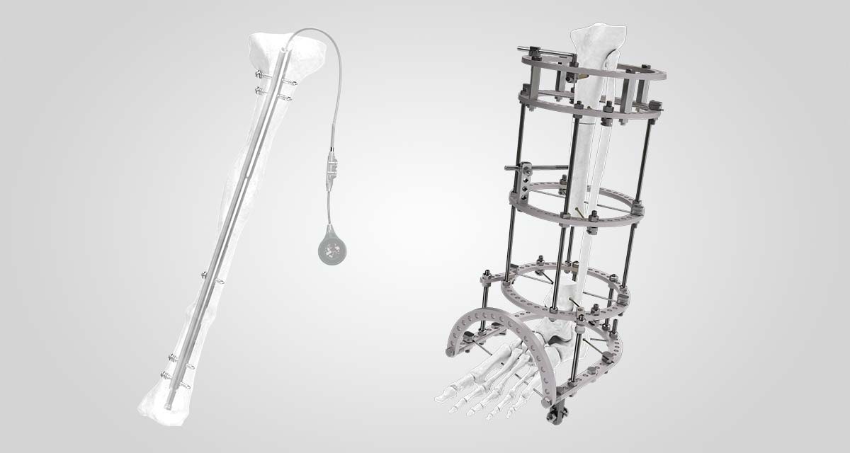
The classic technique described by Prof. Ilizarov utilized an external circular fixator to perform the bone transport. This type of external fixator, also known as the Ilizarov external fixator, is still preferred by some surgeons due to its ability to minimize angulation and rotation movement, which can hinder the bone healing process.(1,13) However, in certain body locations, such as the femur or the humerus, the use of circular external fixation is cumbersome for the patient, making monolateral external fixators or intramedullary bone transport nails more appropriate for those locations.(1,14)
The bone transport technique, albeit effective, requires a lengthy treatment period. All bone segments must be kept stable for the entire time that the new bone is growing and beyond, which means that the external fixator must be used for a long time.(2) To reduce the external fixation time (EFT) and the likelihood of fixator-associated complications, hybrid bone transport techniques were developed.(1)
One of the earlier hybrid bone transport techniques developed was the use of an intramedullary nail after removing the external fixator, in a sequential procedure called Transport And Then Nailing (TATN). It is also possible to perform simultaneous bone transport and internal fixation, with Bone Transport Over a Nail (BTON). In this case, the intramedullary nail maintains alignment and confers stability across the bone defect segment, while the external fixator is used to perform the bone transport.(1) The use of an intramedullary nail adds rigidity to the overall construct and allows for earlier removal of the external fixator, in addition to maintaining alignment.(2)
Bone plates have also been used in hybrid bone transport techniques, being particularly useful in cases of metaphyseal fractures with limited bone stock. In the Plate-Assisted Bone Segment Transport (PABST), a long locking plate is used to maintain alignment and stability in the same way an external fixator would, keeping the proximal and distal bone ends securely in place while the transported bone segment is advanced by the telescoping intramedullary nail.(15-17) This technique may use a temporary external fixator to ensure the proper alignment of the plate; however, this is considered optional.(15) Therefore, the use of plates to stabilize and keep bone alignment can help eliminate use of external fixators and the associated EFT.(16,17) Application of plates are more invasive and associated with high infection rates.
The development of motorized intramedullary nails specifically designed for bone transport allows for an all-internal procedure, without the need for an external fixator or plate, thus avoiding the typical complications associated with external fixators and having the potential of a faster rehabilitation process.(1,2)
The discovery of the bone regeneration potential and the development of the bone transport technique by Prof. Ilizarov constituted a major breakthrough in orthopedic surgery. They remain the foundation upon with new orthopedic devices are created with the goal of improving patient outcomes, by increasing the rate of limb salvage while reducing complications.(1)
This technique plays a crucial role in limb reconstruction, not only addressing bone gap repair but also ensuring the restoration of functional mobility and maintaining the structural integrity of affected limbs.
Conditions Treated with the Bone Transport Technique
Bone segmental loss can be caused by several factors, including trauma, infections (osteomyelitis), bone non-union or malunion, avascular necrosis, or tumors.(12,18) Large bone defects can be difficult to manage, especially when the surrounding soft tissues are compromised.(6,19) These complex cases cannot be addressed with acute correction, limiting the use of techniques like bone grafting.(12) For bone defects of critical size and mangled extremities, amputation is also a treatment option and one that is dependent on the extent of soft tissue injury.(7) However, many patients have a strong preference for limb salvage over amputation, despite the longer treatment and recovery times.(7)
The use of gradual correction is useful not only for addressing large bone defects, but also in cases where there is associated bone deformity or there is a need for simultaneous bone lengthening.(12,20) The unique ability of the bone transport technique to promote bone repair and growth as well as regeneration of the surrounding soft tissue structures (muscles, blood vessels, and nerves) makes it the preferred approach to manage complex limb salvage cases.(12)
This is illustrated in an article by Hosny and Ahmed,(7) in which the authors describe their experience with the bone transport technique to reconstruct complex and neglected war injuries. These patients had been seen in other centers and been offered amputation as treatment, but they chose a second opinion and opted for limb reconstruction. Most patients had good functional and radiological results, emphasizing the importance of bone transport to address complex cases.(7)
Large bone defects are challenging to treat, even when the soft tissue envelope is relatively intact. The soft tissue must be preserved, and the optimal bone regeneration conditions should be established. This includes obtaining an infection-free environment and an ideal vascular bed for the bone to regenerate.(1) Limb salvage cases are aggravated by soft tissue loss, reduced vascularity, and infections.(7,8) The maintenance of an infection-free environment and stable vascular supply is even more important in cases of chronic osteomyelitis, where the infected bone has to be resected and the optimal conditions must be maintained throughout the regeneration process.(21)
The Bone Transport Technique Procedure: Stages and Process
The bone transport technique described by Prof. Ilizarov begins with a corticotomy, or low-energy osteotomy of the bone cortex, that preserves the local blood supply of both the periosteum and the medullary canal.(2,5) There are several techniques to achieve a corticotomy, but prevention of thermal necrosis is the key aspect to consider.(18) Although earlier publications have highlighted the importance of protecting the bone marrow during the corticotomy, later experiments and clinical results have shown that while the endosteum and bone marrow are not indispensable for adequate callus formation, the periosteum is particularly relevant for adequate regenerate formation.(5,13,22)
After the corticotomy, there must be a latency period before the start of the distraction period.(1,5) This provides time for the biologic environment around the corticotomy to start a healing response, which is critical for successful bone regeneration.(18) Starting distraction immediately after the corticotomy can result in slow callus formation; whereas, delaying the start of distraction can cause premature fusion.(22) The usual latency period is 3 to 10 days, but patients with comorbidities, undergoing immunosuppressive therapy, or with infections may need a longer latency period.(14,18) The type of corticotomy also influences the ideal latency period, with more aggressive osteotomy techniques benefitting from a longer latency period.(13)
The distraction period refers to the time when new bone is formed between the widening gap of the osteotomized bone ends. In his experiments, Prof. Ilizarov discovered that a daily distraction rate of 1mm per day was optimal for bone regeneration, while a 0.5mm/day rate often led to premature consolidation and a 2mm/day rate resulted in a poor quality regenerate.(5) The 1mm/day rate achieves the best results when applied in 0.25mm increments, 4 times a day.(18) The increments can be smaller and often produce better results, but such small adjustments are only possible with motorized devices.(5)
As mentioned before, the formation of a good quality regenerate will depend on the mechanical and biological conditions.(18) The mechanical conditions are assured by the rigidity of the construct, while the biological conditions can vary from patient to patient.(13,18) When the distraction ends and the bone segment that is being advanced covers the entire span of the bone defect, there is docking of the contacting bone ends followed by consolidation.1 While the regenerate is consolidating and the bone ends at the docking point are in the process of union, the fixator must be retained.1 The most reliable indicator for consolidation is radiographic evidence of tricortical consolidation on two orthogonal radiographs, although the ability to stand on the treated leg may also be used to assess bone consolidation.(11,18)
One of the main drawbacks of using an external fixator to perform bone transport is the lengthy EFT.(1) Patients can become intolerant to the device, especially during the consolidation phase, when the bone is no longer being distracted. However, premature removal of the device can cause many problems, including fracture, secondary deformity, and non-union.(1,19) To reduce the EFT, hybrid approaches have been developed, with internal and external fixators being applied either sequentially or simultaneously.(1) This allows for adequate bone stabilization and reduced EFT, while improving patient comfort.(1) The use of intramedullary nails can be effectively used for bone transport, allowing early weight-bearing and having low impact on soft-tissues.(23)
Complications and Risks Associated with the Bone Transport Technique
The bone transport technique is not without complications, which can be classified according to their time of onset (immediate versus delayed), if they are related to the surgery or the device, and the anatomical structures involved.(2,18) Surgeons should have enough experience and thorough anatomical knowledge to avoid damage to essential structures during the corticotomy and fixator placement.(2) Patients should be counselled about the possible complications, pain during treatment and length of the treatment.(18)
The most common complication seen during bone transport with external fixation is pin site inflammation and infection.(1,2,10,18,20) This is a problem that can be avoided with adequate post-operative management, including checking if wires have the appropriate tension and providing meticulous pin site care.(19,20,24) If a superficial infection develops, antibiotics should be initiated to prevent progression and eventual deep infection.(20,24)
Muscle contractures and joint stiffness are other possible complications arising from prolonged EFT. They can be prevented with good pre-operative planning, careful positioning of the external fixator, and by following an adequate physical therapy regimen during the distraction and consolidation phases.(1,2,19,25)
The stability of the fixator is essential to prevent axial deviation, which would result in deformity of the regenerate.(2,19) However, the removal of the fixator must be carefully planned, as an early removal may cause instability at the docking site while late removal increases the risk of pin site infections and joint contracture.(1)
The use of internal fixation eliminates complications such as pin site infection and those related to a lengthy EFT, but the constructs are usually not as stable, increasing the risk of axial deviation. To reduce this risk, patients are not allowed to fully weightbear immediately after surgery.(26) The first models of internal lengthening nails relied on patient movement to activate the telescoping mechanism, which means that the distraction rate was done in a relatively uncontrolled way. This contributed to insufficient callus formation in some cases and to extreme pain in others.(27,28) The recently introduced technologies have made huge improvements and the newest models of intramedullary nails for bone transport rely not on patient movements to activate the telescoping motion, but rather on a remotely activated motor.(26) This leads to less pain, although other complications persist, such as nail bending, migration, and bending.(27,28)
As for delayed complications, several authors note that when the bone defect is larger than 8cm, there is an increased risk of fracture, either of the regenerate or at the docking site.(1,2,6,10)
Bone Transport Technique Outcomes and Success Rate
The management of complex bone defects often requires a personalized approach, since it depends on various factors, including the patient status, the defect size, the quality of the surrounding soft tissues, the presence of deformity or length discrepancy, and the experience of the surgeon.(11)
The fixation time depends on the length of bone defect and how well the regenerate consolidates.(13) This means that it is not uncommon for patients to have an external fixator in place for 4 to 12 months, although this time can go up to 28 months in complex cases with large bone defects.(13,29) Before removing the external fixator, bone consolidation should be assessed through radiographic imaging and through clinical assessment.(11,13) The bone regenerate is assessed regarding its homogeneity, density, and neocorticalization. If no pain or inflammation develops, then the external frame can be safely removed.(13)
The bone transport technique is effective in promoting bone healing across large bone defects and in eliminating long-standing bone infections and non-unions, but the lengthy treatment and recovery are burdensome for patients.(6,12) Patient compliance during the bone transport treatment is a key factor to prevent complications and improve outcomes, highlighting the importance of patient selection and counselling.(1,18)
Most patients who undergo bone transport have good functional outcomes, retaining limb functionality and bone quality. Some patients have exhausted other treatment modalities and face amputation, but with careful planning, bone transport is still a viable choice for limb salvage, avoiding amputation.(7,8)
Amputation rates are low in studies evaluating the use of bone transport, with most amputations being requested by the patients.(6) This highlights not only the effectiveness of the technique but the patient burden, which in many cases is caused by the EFT. Therefore, future developments are focusing on reducing patient burden by developing post-surgery protocols focusing on avoiding common complications and reducing EFT time with all-internal bone fixation devices.(1,24,30)
Conclusions
The bone transport technique remains an indispensable tool in limb reconstruction surgery, offering hope to patients facing limb amputation. As innovations like motorized nails and hybrid fixation systems continue to evolve, the future of bone defect reconstruction promises improved outcomes and reduced patient burden.
References
- Seng DWR, Oh C-W. Critical size bone defects managed with modern techniques of bone transport: An update. Injury. 2024/03/01/ 2024;55(3):111341. doi:https://doi.org/10.1016/j.injury.2024.111341
- Chimutengwende-Gordon M, Mbogo A, Khan W, Wilkes R. Limb reconstruction after traumatic bone loss. Injury. 2017/02/01/ 2017;48(2):206-213. doi:https://doi.org/10.1016/j.injury.2013.11.022
- C R, S J, Tabrizi M. Distraction Osteogenesis and Its Challenges in Bone Regeneration. InTech; 2012.
- 4. Li J, Li M, Wang W, Li B, Liu L. Evolution and development of ilizarov technique in the treatment of infected long bone nonunion with or without bone defects. Orthopaedic Surgery. 2022/05// 2022;14(5):824-830. doi:10.1111/os.13218
- Ilizarov GA. Clinical Application of the Tension–Stress Effect for Limb Lengthening. Clinical Orthopaedics and Related Research®. 1990;250
- Papakostidis C, Bhandari M, Giannoudis PV. Distraction osteogenesis in the treatment of long bone defects of the lower limbs: Effectiveness, complications and clinical results; a systematic review and meta-analysis. The Bone & Joint Journal. 2013/12// 2013;95-B(12):1673-1680. doi:10.1302/0301-620X.95B12.32385
- 7Hosny GA, Ahmed A-SA-A. Neglected war injuries: Reconstruction versus amputation. Injury. 2023/12/01/ 2023;54(12):111085. doi:https://doi.org/10.1016/j.injury.2023.111085
- Farrelly E, Tarapore R, Lindsey S, Wieland MD. Management of the mangled extremity. Surgical Clinics of North America. 2024/04// 2024;104(2):385-404. doi:10.1016/j.suc.2023.10.006
- Weinlein CJ. Delayed Union and Nonunion of Fractures. In: Azar FM, Beaty JH, Canale ST, eds. Campbell’s operative orthopaedics. 14th edition. ed. Elsevier; 2021.
- Aktuglu K, Erol K, Vahabi A. Ilizarov bone transport and treatment of critical-sized tibial bone defects: a narrative review. J Orthop Traumatol. 2019/12// 2019;20(1):22. doi:10.1186/s10195-019-0527-1
- El-Rosasy M, Mahmoud A, El-Gebaly O, Rodriguez–Collazo E, Thione A. Definition of bone transport from an orthoplastic perspective. International Journal of Orthoplastic Surgery. 2019/06/20/ 2019;2(2):62-71. doi:10.29337/ijops.33
- Rozbruch SR, Pugsley JS, Fragomen AT, Ilizarov S. Repair of tibial nonunions and bone defects with the taylor spatial frame. Journal of Orthopaedic Trauma. 2008/02// 2008;22(2):88-95. doi:10.1097/BOT.0b013e318162ab49
- Catagni MA, Guerreschi F, Lovisetti L. Distraction osteogenesis for bone repair in the 21st century: Lessons learned. Injury. 2011/06// 2011;42(6):580-586. doi:10.1016/j.injury.2011.04.004
- Iacobellis C, Berizzi A, Aldegheri R. Bone transport using the Ilizarov method: a review of complications in 100 consecutive cases. Strategies in Trauma and Limb Reconstruction. 2010/04/30/ 2010;5(1):17-22. doi:10.1007/s11751-010-0085-9
- Olesen UK, Nygaard T, Prince DE, et al. Plate-assisted Bone Segment Transport With Motorized Lengthening Nails and Locking Plates: A Technique to Treat Femoral and Tibial Bone Defects. J Am Acad Orthop Surg Glob Res Rev. Aug 2019;3(8):e064. doi:10.5435/JAAOSGlobal-D-19-00064
- Eldesouqi AA, Yau RCH, Ho W-YK, Lam Y-L. Plate-assisted bone segment transport: Novel application on distal tibia defect after tumour resection. A case report. International Journal of Surgery Case Reports. 2021/07/01/ 2021;84:106079. doi:https://doi.org/10.1016/j.ijscr.2021.106079
- Gardner MP, Beason AM. Plate-Assisted Bone Segment Transport Versus Precice Bone Transport Nail. Journal of Orthopaedic Trauma. 2021;35
- Millonig K, Hutchinson B. Management of osseous defects in the tibia. Clinics in Podiatric Medicine and Surgery. 2021/01// 2021;38(1):111-116. doi:10.1016/j.cpm.2020.09.006
- Schnack LL, Oexeman S, Rodriguez-Collazo ER. Management of Osseous Defects of the Tibia Utilizing Orthofix Hexapod Circular External Fixator: A Technique Guide—An Orthoplastic Approach for Combined Soft Tissue and Osseous Defects. Clinics in Podiatric Medicine and Surgery. 2021/01/01/ 2021;38(1, Supplement):e44-e58. doi:https://doi.org/10.1016/j.cpm.2021.09.003
- Tellisi N, Fragomen AT, Ilizarov S, Rozbruch SR. Limb salvage reconstruction of the ankle with fusion and simultaneous tibial lengthening using the ilizarov/taylor spatial frame. HSS Journal®: The Musculoskeletal Journal of Hospital for Special Surgery. 2008/02// 2008;4(1):32-42. doi:10.1007/s11420-007-9073-0
- McNally M, Ferguson J, Kugan R, Stubbs D. Ilizarov Treatment Protocols in the Management of Infected Nonunion of the Tibia. Journal of Orthopaedic Trauma. 2017;31
- Kojimoto H, Yasui N, Goto T, Matsuda S, Shimomura Y. Bone lengthening in rabbits by callus distraction. The role of periosteum and endosteum. The Journal of Bone and Joint Surgery British volume. 1988/08// 1988;70-B(4):543-549. doi:10.1302/0301-620X.70B4.3403595
- Carrión Martínez J, Cámara Baeza MA, Durán Morell A, Mas PC, González Gil AB. Treatment of a tibial bone defect with a motorized intramedullary bone transport nail: A case review with 32 months follow up. Trauma Case Reports. 2022/12/01/ 2022;42:100718. doi:https://doi.org/10.1016/j.tcr.2022.100718
- Marais LC. Prevention and management of external fixator pin track sepsis. Strategies in Trauma and Limb Reconstruction. 2012/08/31/ 2012;7(2):67-72. doi:10.1007/s11751-012-0139-2
- Al Shahrani AA, Tedla JS, Ahmad I. Effectiveness of Ilizarov frame fixation on functional outcome in aseptic tibial non-union cases at Abha, Kingdom of Saudi Arabia: An experimental study. Journal of Taibah University Medical Sciences. 2015/06// 2015;10(2):216-221. doi:10.1016/j.jtumed.2014.09.002
- Schultz BJ, McLaurin TM, Leucht P. Treatment of Segmental Bone Defects Biology and Treatment Options. Bull Hosp Jt Dis (2013). Mar 2022;80(1):53-64.
- Singh S, Lahiri A, Iqbal M. The results of limb lengthening by callus distraction using an extending intramedullary nail (fitbone) in non-traumatic disorders. Article. Journal of Bone and Joint Surgery – Series B. 2006;88(7):938-942. doi:10.1302/0301-620X.88B7.17618
- Krieg AH, Speth BM, Foster BK. Leg lengthening with a motorized nail in adolescents: an alternative to external fixators? Clin Orthop Relat Res. Jan 2008;466(1):189-97. doi:10.1007/s11999-007-0040-3
- Yang Z, Tao H, Ye Z, Jin L, Lin N, Yang D. Bone transport for reconstruction of large bone defects after tibial tumor resection: a report of five cases. J Int Med Res. Aug 2018;46(8):3219-3225. doi:10.1177/0300060518774992
- Jiang L, Mendame Ehya RE. Effectiveness of a collaborative nursing care model for the treatment of patients with diabetic foot disease by transverse tibial bone transport technique: a pilot study. Journal of PeriAnesthesia Nursing. 2020/02// 2020;35(1):60-66. doi:10.1016/j.jopan.2019.06.009


